Diabetic eye disease is the leading cause of blindness in the world. If you or a loved one has diabetes, it is so important to have a yearly comprehensive eye exam. Call us today for your appointment!
Always ensure to wear the sunglasses to protect your delicate eyes against blazing sun this summer!
Are you ready to enjoy life without the hassle of glasses or contacts? Take the first step by calling Eye Care Center today at 01 747078/9 for your LASIK evaluation!
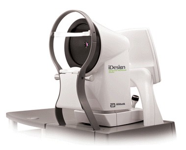
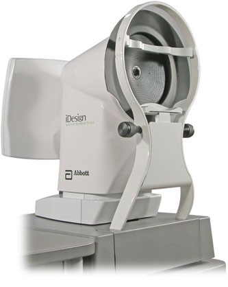
The one and ONLY iDesign Advanced WaveScan Studio System in Lebanon from Abbott Medical Optics allows your physician to customize the procedure just for you, making possibilities realities. Start seeing life to the MAX!
Tour ECCand Enjoy the Latest in Ophthalmic Diagnostic and Treatment Equipment.
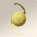
Multifocal IOLsare now used in up to 40% of cataract surgery in Europe and the US. Dr. Pierre Mardelli was the first surgeon to have used it in Lebanon and the Middle East back in 2004. Come benefit from our years of experience and let us show you the difference.
BOTOX®Cosmetic is injected into muscles and used to improve the look of moderate to severe frown lines between the eyebrows (glabellar lines) and around the eyelids for a short period of time (temporary). At ECC we aim for a more "natural look".
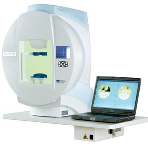
Glaucoma is the second leading cause of blindness. ECC acquired the latest Visual Field Analyzer from Haag Streit and OCT from Optovue. Both are vital for earlier diagnosis and follow-up of this blinding disease.
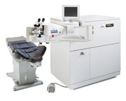
The newest generation excimer laser from AMO: The VISX S4 with the iDesign Advanced Wavescan Studio (TM). This is the first system of its kind in Lebanon and the Middle East. Go ahead and call us for more info.
Custom LASIKwith the iDesign Wavescan Studio System combines two exciting technologies to reshape the cornea to reduce or eliminate visual distortions. This technology has the potential to improve how much and how well you can see as well as reduce the risks of post-LASIK complications.
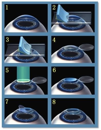
LASIKis a refractive surgical procedure that corrects your vision, thus lessening your dependence on any corrective devices such as contact lenses or eyeglasses. At ECC we strive to offer you the latest technology such as Custom LASIK using the iDesign Wavescan.
With the new aquisition of the latest generation excimer laser, ECC is setting the bar ever higher. We strive to provide our patients with the best customer-care experience and quality of vision available today.
NEWS & ARTICLES
Age-Related Macular Degeneration (AMD)
The macula makes up only a small part of the retina, yet it is much more sensitive to detail than the rest of the retina (called the peripheral retina). The macula is what allows you to thread a needle, read small print, and read street signs. The peripheral retina gives you side (or peripheral) vision. If someone is standing off to one side of your vision, your peripheral retina helps you know that person is there by allowing you to see their general shape.
Many older people develop macular degeneration as part of the body's natural aging process. There are different kinds of macular problems, but the most common is age-related macular degeneration.
One symptom of macular degeneration is dark areas in your central vision.
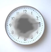
With macular degeneration, you may have symptoms such as blurriness, dark areas or distortion in your central vision, and perhaps permanent loss of your central vision. It usually does not affect your side, or peripheral vision. For example, with advanced macular degeneration, you could see the outline of a clock, yet may not be able to see the hands of the clock to tell what time it is.
Causes of macular degeneration include the formation of deposits called drusen under the retina, and in some cases, the growth of abnormal blood vessels under the retina. With or without treatment, macular degeneration alone almost never causes total blindness. People with more advanced cases of macular degeneration continue to have useful vision using their side, or peripheral vision. In many cases, macular degeneration's impact on your vision can be minimal.
When macular degeneration does lead to loss of vision, it usually begins in just one eye, though it may affect the other eye later.
Many people are not aware that they have macular degeneration until they have a noticeable vision problem or until it is detected during an eye examination.
Types of macular degeneration: dry macular degeneration and wet macular degeneration
There are two types of macular degeneration:
Dry, or atrophic, macular degeneration (also called non-neovascular macular degeneration) with drusen
Most people who have macular degeneration have the dry form. This condition is caused by aging and thinning of the tissues of the macula. Macular degeneration usually begins when tiny yellow or white pieces of fatty protein called drusen form under the retina. Eventually, the macula may become thinner and stop working properly.
With dry macular degeneration, vision loss is usually gradual. People who develop dry macular degeneration must carefully and constantly monitor their central vision. If you notice any changes in your vision, you should tell your ophthalmologist (Eye M.D.) right away, as the dry form can change into the more damaging form of macular degeneration called wet (exudative) macular degeneration. While there is no medication or treatment for dry macular degeneration, some people may benefit from a vitamin therapy regimen for dry macular degeneration.
Using an Amsler grid to test for macular degeneration If you have been diagnosed with dry macular degeneration, you should use a chart called an Amsler grid every day to monitor your vision, as dry macular degeneration can change into the more damaging wet form.
To use the Amsler grid, wear your reading glasses and hold the grid 12 to 15 inches away from your face in good light.
Cover one eye.
Look directly at the center dot with the uncovered eye and keep your eye focused on it.
While looking directly at the center dot, note whether all lines of the grid are straight or if any areas are distorted, blurry or dark.
Repeat this procedure with the other eye.
If any area of the grid looks wavy, blurred or dark, contact your ophthalmologist.
If you detect any changes when looking at the grid, you should notify your ophthalmologist immediately.
Wet, or exudative, macular degeneration (also called neovascular macular degeneration)
About 10 percent of people who have macular degeneration have the wet form, but it can cause more damage to your central or detail vision than the dry form.
Wet macular degeneration occurs when abnormal blood vessels begin to grow underneath the retina. This blood vessel growth is called choroidal neovascularization (CNV) because these vessels grow from the layer under the retina called the choroid. These new blood vessels may leak fluid or blood, blurring or distorting central vision. Vision loss from this form of macular degeneration may be faster and more noticeable than that from dry macular degeneration.
The longer these abnormal vessels leak or grow, the more risk you have of losing more of your detailed vision. Also, if abnormal blood vessel growth happens in one eye, there is a risk that it will occur in the other eye. The earlier that wet macular degeneration is diagnosed and treated, the better chance you have of preserving some or much of your central vision. That is why it is so important that you and your ophthalmologist monitor your vision in each eye carefully.
Dry macular degeneration signs and symptoms
Blurry distance and/or reading vision
Need for increasingly bright light to see up close
Colors appear less vivid or bright
Hazy vision
Difficulty seeing when going from bright light to low light (such as entering a dimly lit room from the bright outdoors)
Trouble or inability to recognize people's faces
Blank or blurry spot in your central vision
Dry macular degeneration can affect one or both eyes. You may not notice vision changes if only one eye is affected, as your unaffected eye will compensate for vision loss in the other eye.
Wet macular degeneration signs and symptoms
Distorted vision — straight lines will appear bent, crooked or irregular
Dark gray spots or blank spots in your vision
Loss of central vision
Size of objects may appear different for each eye
Colors lose their brightness; colors do not look the same for each eye
Wet macular degeneration symptoms usually appear and get worse fairly quickly.
Oxidative stress and macular degeneration
Our bodies constantly react with the oxygen in our environment. Over our lifetimes, as a result of this activity, our bodies produce tiny molecules called free radicals. These free radicals affect our cells, sometimes damaging them. This is called oxidative stress and is thought to play a major role in how macular degeneration develops. Approximately 1 in 3 Caucasians have genetic changes that make them more prone to damage from oxidative stress, which can lead to macular degeneration.
Macular degeneration in families
Heredity is another risk factor for macular degeneration. People who have a close family member with the disease have a greater chance of developing macular degeneration themselves.
Inflammation and macular degeneration
Some studies have shown that inflammation (swelling of the body’s tissues) may play a role in macular degeneration development. Inflammation is the way the body’s immune system fights off infection or other things it considers “invaders.” But an overactive immune system with its associated inflammation may be a risk factor for macular degeneration.
Smoking, high blood pressure and abnormal cholesterol and macular degeneration
Smoking and high blood pressure are associated with the wet form of macular degeneration. Research also suggests there may be a link between being obese and having early or intermediate-stage macular degeneration develop into the advanced (wet) form.
Another risk factor for developing macular degeneration may include having abnormal cholesterol levels or having high blood pressure (called hypertension).
Dry macular degeneration: detection with an ophthalmoscope
To check for macular degeneration, your eye doctor will dilate (widen) your pupils using eyedrops and examine your eyes with an ophthalmoscope, a device that allows him or her to see the retina and other areas at the back of the eye. If macular degeneration is detected, your doctor may have you use an Amsler grid to check for macular degeneration symptoms such as wavy, blurry or dark areas in your vision.
Wet macular degeneration: detection with fluorescein angiography and optical coherence tomography
If your ophthalmologist suspects you may have the wet form of macular degeneration, he or she will take special photographs of your eye with fluorescein angiography and optical coherence tomography (OCT). OCT scanning is a sophisticated and exact tool that detects abnormal blood vessels by creating a special picture of your macula.
During fluorescein angiography, a fluorescein dye is injected into a vein in your arm. The dye travels throughout the body, including your eyes. Photographs are taken of your eye as the dye passes through the retinal blood vessels. Abnormal areas will be highlighted by the dye, showing your doctor whether wet macular degeneration treatment is possible and, if so, where to treat the abnormal vessels.
The Age-Related Eye Disease Study (AREDS) showed that among people at high risk for developing late-stage, or wet, macular degeneration (such as those who have large amounts of drusen or who have significant vision loss in at least one eye), taking a dietary supplement of vitamin C, vitamin E and beta carotene, along with zinc, lowered the risk of macular degeneration progressing to advanced stages by about 25 percent. The daily supplements also reduced the risk of vision loss for those at risk by about 19 percent. The supplements did not appear to provide a benefit for people with minimal macular degeneration or people without evidence of the disease during the course of the study.
Following is the nutrient supplementation shown to be beneficial in lowering the risk of macular degeneration progressing to advanced stages:
• Vitamin C – 500 mg
• Vitamin E – 400 IU
• Beta carotene – 15 mg (25,000 IU)
• Zinc oxide – 80 mg
• Copper (as cupric oxide) – 2 mg (to prevent copper deficiency, which may be associated with taking high amounts of zinc)
Another large study in women showed a benefit from taking folic acid and vitamins B6 and B12. And a large study evaluating the possible benefits of lutein and fish oil (omega-3) is ongoing. Other studies have shown that eating dark leafy greens, yellow, orange and other colorful fruits and vegetables, rich in lutein and zeaxanthin, may reduce your risk for developing macular degeneration.
These vitamins and minerals are recommended in specific daily amounts in addition to a healthy, balanced diet. Some people may not wish to take large doses of antioxidants or zinc because of medical reasons. Beta carotene has been shown to increase the risk of lung cancer in smokers or recent past smokers, so this supplement should not be used by people who currently smoke or recently quit smoking.
It is very important to remember that vitamin supplements are not a cure for macular degeneration, nor will they give you back vision that you may have already lost from the disease. However, specific amounts of these supplements do play a key role in helping some people at high risk for developing advanced (wet) AMD to maintain their vision.
Talk with your ophthalmologist to find out if you are at risk for developing advanced macular degeneration, and to learn if supplements are recommended for you.
Wet macular degeneration treatment
Treating the wet form of macular degeneration may involve the use of anti-VEGF treatment, thermal laser treatment or photodynamic therapy (PDT). Treatment of wet macular degeneration generally reduces—but does not eliminate-- the risk of severe vision loss.
Anti-VEGF medication injection treatments for wet macular degeneration
A common way to treat wet macular degeneration targets a specific chemical in your body that causes abnormal blood vessels to grow under the retina. That chemical is called vascular endothelial growth factor, or VEGF. Several new drug treatments (called anti-VEGF drugs) have been developed for wet AMD that can block the trouble-causing VEGF. Blocking VEGF reduces the growth of abnormal blood vessels, slows their leakage, helps to slow vision loss, and in some cases improves vision.
Your ophthalmologist administers the anti-VEGF drug directly to your eye in an outpatient procedure. Before the procedure, your ophthalmologist will clean your eye to prevent infection and will use an anesthetic to numb your eye with a very fine needle. You may receive multiple anti-VEGF injections over the course of many months. Repeat anti-VEGF treatments are often needed for continued benefit.
In some cases, your ophthalmologist may recommend combining anti-VEGF treatment with other therapies. The treatment that’s right for you will depend on the specific condition of your macular degeneration.
Laser treatment for wet macular degeneration
Another form of treatment for wet AMD is with thermal laser therapy. Laser treatment is usually done as an outpatient procedure in the doctor’s office or at the hospital.
The laser beam in this procedure is a high-energy, focused beam of light that produces a small burn when it hits the area of the retina to be treated. This destroys the abnormal blood vessels, preventing further leakage, bleeding and growth.
Following laser treatment, vision may be more blurred than before treatment, but often it will stabilize within a few weeks. A scar forms where the treatment occurred, creating a permanent blind spot that might be noticeable in your field of vision.
Usually the abnormal blood vessels are destroyed by later treatment. However, it is likely that 50 percent of patients with wet macular degeneration who receive this laser procedure will need a re-treatment within three to five years. You may be instructed to use the Amsler grid daily to monitor your vision for signs of change.
Photodynamic therapy (PDT)
In some cases, a type of treatment for wet macular degeneration called photodynamic therapy, or PDT, may be an option. This therapy uses a combination of a light-activated drug called a photosensitizer and a special low-power, or cool, laser to treat wet macular degeneration right at the center of the macula.
This procedure is done on an outpatient basis, usually in an ophthalmologist’s office. The photosensitive drug is injected into a vein in your arm, where it travels through the body, including the abnormal vessels behind the central macula. The low-power laser light is targeted directly on the abnormal vessels, activating the drug, which causes damage specifically to those unwanted blood vessels.
After PDT, the abnormal blood vessels may reopen, so multiple treatments may be required.
What happens when macular degeneration cannot be treated?
It is important to remember that only about 10 percent of all macular degeneration cases are exudative, or wet form, and about 75 percent of these cases cannot be treated. People with wet or dry macular degeneration symptoms who cannot be treated will not become blind, as they will still have peripheral, or side, vision.
If you have untreatable macular degeneration, you can make the most of your remaining vision by learning to “see again” with the vision you do have and with the help of special low-vision rehabilitation, devices and services. People with low vision can learn new strategies to accomplish daily activities. These skills, including mastering new techniques and devices, help people with advanced AMD regain their confidence and live independently despite loss of central vision.
While there is little that can be done to improve the eyesight of someone who has AMD, with early detection, the rate of vision loss can be slowed. The keys to slowing vision loss are to understand macular degeneration, monitor your symptoms and visit your ophthalmologist regularly to test your vision. Even with macular degeneration, you can still maintain an enjoyable lifestyle.